
Hysteroscopic Endometrial Ablation/Resection of submucous uterine fibroids
/The Resectoscope is an instrument very similar to the one used to treat the enlarged prostate in the male. It is a telescope that is 10mm in diameter. The telescope can be used with 2 separate small attachments. One is an insulated loop, which cuts and coagulates at the same time, and the other is a ball, which literally rolls up and down the intrauterine surface. People with heavy periods may elect to have the endometrium “cored” out much the same way as an apple is “cored” out. This is analogy only!

To do this, the endometrium (lining of the uterus) is firstly thinned down, using a variety of medications. The medications used are only short term – about 6 weeks is usually enough. By thinning the lining of the womb down, I can get a much deeper “cut” into the uterine muscle without having my view occluded by unnecessary chips, or pieces of resected endometrium.

Once the endometrium is thinned, the patient is admitted, she is put to sleep, the hysteroscope is inserted under vision from a monitor, into the uterine cavity, and until recently my chosen method was to use just the loop with the diathermy attached, using both cutting and coagulation diathermy power, as need be .I often use a blend of both cutting and coagulation.
The loop resects the endometrium and also goes deeper into the muscle of the uterus. This is particularly useful with adenomyosis. The tissue can also be sent for histology to confirm that there is no cancer or pre-cancer present. The procedure takes about 45 minutes to perform. It is done under a general anaesthetic. I send my patients home the same day and generally patients suffer only mild crampy period like pain – enough to take just Panadol or perhaps some Nurofen.
Recently, I have begun inserting a Mirena device into the uterus, after the resection, to try to prevent any regrowth of endometrium from deeper areas of the remaining muscle which may contain remnants of adenomyosis, which could grow back and resurface the endometrium. This works well.
Even though there are very few complications from this operation, those complications can indeed be life threatening. The major risk of this procedure is that perforation of the uterus may occur, and that damage to the blood vessels and bowel. Should any perforation to the uterus occur, an emergency laparotomy(large cut in the abdomen), and hysterectomy would need to be performed. I warn my patients about this prior to the surgery. This of course, is a rare event. In the last 28 years since I have been in private practice, this has occurred only twice out of many hundreds of operations I have performed.
The roller ball is also another attachment that can be put in through the hysteroscope. This is certainly safer in the hands of the less experience operator, but I feel it is very difficult to know how deep to go with the diathermy (burn),when the ball is used alone. In my hands, I do not feel the success rate is as high, when the roller ball is used alone. I therefore reserve the use of the roller ball for the patient who does not wish to have the Mirena device inserted after her resection has been done. The roller ball is a bit like putting the finishing operative touches on the lining of the wound, after the major work has been done by the endometrial resection loop itself.
The original operations to treat menorrhagia in the United States ,and indeed Australia through the hysteroscope were done using a laser, which was inserted though the hysteroscope. In a similar way that a spray painter would paint a car, the laser beam was “painted” across the internal cavity of the uterus thus vaporising the endometrium.
Although the laser was successful, it was and is, an extremely expensive way to remove endometrium and few facilities offer this anymore.
Other ways to ablate the Endometrium
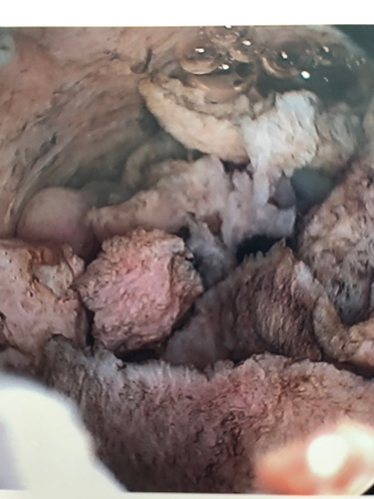
For a few years I did replace the endometrial resection procedure with a new device, which involved a new technique. This involved inserting a balloon into the endometrial cavity and keeping that in the endometrial cavity for a full 10 minutes, once the “operating temperature” was reached , the endometrium literally boiled away. Hysteroscopic photos taken before and after the procedure showed that the endometrium took on a brownish rather than a reddish tinge.
The balloon device was completely disposable and a record was kept of the temperature using a computerized system. It was indeed a safer system than endometrial ablation in the hands of less experienced operators. It did not involve any moving parts and there was much less chance for human error. Certainly, the device could not cut through the uterine cavity, because undue force was used. For the inexperienced surgeon, this was a safer option, BUT the results were not as good, and I therefore reverted back to the Resectoscope on which I trained.
Postoperative Symptoms
The success rate of endometrial ablation runs at about 90% “great improvement” in period symptoms, if a Mirena device is inserted at the end of the procedure. There is a small failure rate, and this usually occurs with severe adenomyosis, where the disease affects the outer layer of the uterine muscle.
It is my experience that this procedure of endometrial resection fails due to either inexperience on the part of the operator – not wanting to go to deep for fear of perforation, or more likely to undiagnosed severe adenomyosis.
As will be mentioned later, adenomyosis is where the endometrium grows into the muscle of the uterus and is therefore impossible to resect completely. It is impossible to ablate it, it is impossible to destroy the roller ball, it is impossible to boil the whole of the endometrium away as well. The endometrium in the muscle then resurfaces from the depths of the muscle and in time, often as short as a year, the patient’s periods can be just as heavy as before the operation. Putting the Mirena in, as previously mentioned, helps reduce that risk.
Finally, it is imperative that before any of these procedures are performed, that the patient is seen and assessed gynaecologically with a vaginal examination or vaginal ultrasound scan probe, a Pap smear and assessment of any prolapse that may be present. If a severe prolapse is present and the patient has finished her family, she may indeed be better off having a vaginal hysterectomy. It is mandatory to obtain a sample of the endometrium before operating, and my preference is to perform a hysteroscopy with a biopsy that, so that I can actually visualize the endometrial cavity. I can also then take tissue and send it to the pathologist, so that I know before the operation starts that the patient does not have a cancer. Similarly, by doing this, we exclude the relatively common finding of an endometrial polyp or even fibroids on stalks (pedunculated fibroids), which may simply be resected by themselves ,thus restoring periods to normal.
It is important here to also mention endometriosis! In the early days, many hysteroscopists resected the endometrium, only to find later, that there was an explosion of the patient’s endometriosis. In other words, they had failed to diagnose the endometriosis, which had been present there in the first place, along with the adenomyosis. If there is any suggestion of endometriosis being present it is important to laparoscope the patient at the time of the initial workup. I perform a hysteroscopy and a laparoscopy combined in many cases. That is not to say that the laparoscopic resection of endometriosis cannot be performed at the same time as an endometrial ablation. It can, and I often do first of all, cut away endometriosis, then go below and resect the endometrium.
For further information I refer you to the brochure which I wrote back way back in 1990. The purpose of this brochure was very explicit . I wanted to explain to patients the risks of this procedure in no uncertain terms. When I came back from the United Kingdom in 1990, I had just witnessed my English Consultant, Mr Lloyd Rankin (now retired) teach himself, under the direction of a famous French hysteroscopist how to perform the operation. I saw a few side effects/complications in the hospital I was working at, but more importantly heard about numerous “cowboys” performing the procedure and wanted to make sure my patients were fully informed about these risks. I also wanted to fully inform my older colleagues that I actually taught in Australia, of these risks.
Perforation of the uterus at the time of hysteroscopy is rare. Note that a hysteroscopy is performed using a camera and a large video screen. If the hysteroscope is inserted under “direct vision” (as I do) and the hysteroscopy is being done purely for diagnostic purposes – that is just to have a look and see to confirm or deny the diagnosis of polyps, fibroids or a uterine septum, it is highly unlikely any perforation will occur. Should it occur however, a laparoscopy may need to be undertaken to confirm that no damage has been done to the bowel or blood vessels. Again, perforation is a very rare event. The chance of perforating the uterus is slightly higher when an operating hysteroscopy is undertaken – that is when instruments are inserted down an operating channel, to remove polyps, resect fibroids, “core” out the endometrium as outlined above in the endometrial resection operation.
In these rare cases, because surgical perforation of the uterus will have occurred, a laparotomy or formal abdominal incision may need to be made to ascertain whether any damage to bowl, bladder or blood vessels has occurred.
In my experience of 29 years I have in fact perforated the uterus twice. Once was my 10th operation in England, where I thought I knew it all, and the second time was when I had performed over 400 procedures.
In the first case I needed to perform an emergency hysterectomy. In the latter I did not.